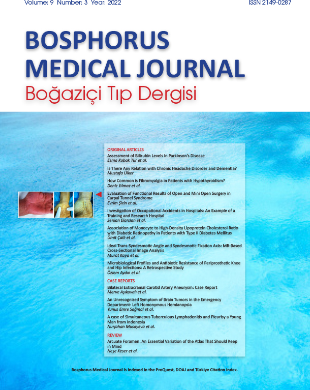
Volume: 9 Issue: 3 - 2022
| BRIEF REPORT | |
| 1. | Frontmatters Pages I - VIII |
| ORIGINAL RESEARCH | |
| 2. | Assessment of Bilirubin Levels in Parkinsons Disease Esma Kobak Tur, Buse Çağla Arı doi: 10.14744/bmj.2022.14227 Pages 145 - 150 INTRODUCTION: Reaktif oksijen radikallerinin aşırı üretimi oksidatif stresi tetikler. Bilirubin oksidasyonu diğer birçok antioksidandan daha güçlü bir şekilde bastırır. Araştırmacılar, Parkinson hastalığı patofizyolojisine büyük önem verdiklerinden, hastalık üzerindeki etkisini incelemişlerdir. Parkinson hastalığı ile antioksidan durum arasındaki ilişki tam olarak araştırılmamıştır. Bu çalışmanın amacı, Parkinson hastalarının serum bilirubin düzeyleri ile demografik ve klinik özellikleri arasındaki ilişkiyi değerlendirmektir. METHODS: Çalışmaya toplam 289 kişi dahil edildi. Serum total, direkt ve indirekt bilirubin düzeyleri demografik ve klinik özelliklerle karşılaştırıldı. Hastalığın şiddeti modifiye Hoehn&Yahr Evreleme Ölçeği ile, klinik özellikleri ise Birleşik Parkinson Hastalığı Derecelendirme Ölçeği ile belirlendi. Hastalar, klinik özelliklerine göre tremor dominant tip, postüral instabilite ve yürüme zorluğu tipi, karışık tip olarak üç motor alt gruba ayrıldı. Modifiye Hoehn&Yahr Evreleme Ölçeği evrelerine göre ise erken (≤2) ve ileri (>2) evre olarak ayrıldı. RESULTS: Çalışma, 189 hasta ve 100 sağlıklı kontrolden oluştu. Yaş ayarlandıktan sonra Parkinson hastalarında indirekt bilirubin düzeyleri anlamlı olarak daha yüksek saptandı (p=0,024). Erkek hastalarda kadın hastalardan daha yüksek bilirubin düzeyleri (direkt, indirekt ve total bilirubin) vardı (p<0,05). Parkinson hastalarının motor alt tipleri arasında ve Parkinson hastalığı evrelerinde bilirubin düzeyleri benzerdi (p>0,05). DISCUSSION AND CONCLUSION: Bilirubin, Parkinson hastalığında dopaminerjik eksikliği gösteren önemli bir antioksidan belirteçtir. Artan bilirubin seviyeleri, Parkinson hastalığının ilerlemesi sırasında ortaya çıkan oksidatif strese karşı geliştirilmiş bir yanıt olabilir. |
| 3. | Is There Any Relation with Chronic Headache Disorder and Dementia? Mustafa Ülker doi: 10.14744/bmj.2022.13471 Pages 151 - 155 INTRODUCTION: Headache disorders can be reasonably speculated to be associated with the increased risk of dementia. However, the current evidence from longitudinal studies linking headache disorders to dementia is scarce, and study populations are often too small to detect clinically relevant associations. In this study, we investigated the incidence of chronic headache history in the dementia population in comparison with the age-matched non-demented control group. METHODS: A total of 122 dementia patients and 114 age-matched non-demented people admitted to outpatient clinic for any complaints other than headache in dates between January 2022 and June 2022 included in this study. Dementia patients who were classified as Alzheimer disease, vascular dementia, mixed dementia, frontotemporal dementia, and Parkinson-related dementia were selected. Mini-mental test score (MMSE) was used to assess the severity of the cognitive impairment. Headache subtypes were classified as migraine headache, episodic tension type headache, and chronic tension-type headache according to International Headache Classification. We investigated and compare the frequency of chronic headache disorders in between two groups. We also aimed to investigate the types of the headaches in demented people and to search the difference from the general population. RESULTS: We could not find any difference statistically between the patient and control groups for the frequency of chronic headache disorder (p=0.568) and between headache subtypes and MMSE (p=0.669). We also examine the relationship between dementia type and subtypes of chronic headache in patient group and we could not find any relationship between this (p=0.070). DISCUSSION AND CONCLUSION: In our small population retrospective study, we could not detect increased chronic headache history in demented people other than non-demented and could not find any relation between chronic headache disorders and dementia. |
| 4. | How Common is Fibromyalgia in Patients with Hypothyroidism? Deniz Yılmaz, Betül Erişmiş, Işıl Üstün, Hakan Koçoğlu, Cemal Bes doi: 10.14744/bmj.2022.60252 Pages 156 - 160 INTRODUCTION: Fibromyalgia (FM) is the most common cause of chronic generalized musculoskeletal pain. The etiology and the pathophysiology are still not clear but there are some studies that elucidate relationship between FM and thyroid diseases. The aim of this study was to determine the frequency of FM in patients with hypothyroidism. METHODS: This is a cross-sectional, single-center, and prospective study from Bakırkoy Dr Sadi Konuk Training and Research Hospital. A total of 180 patients who were applied to internal medicine outpatient clinics included in the study and the patients who described the generalized musculoskeletal pain were consulted to the physical medicine and rehabilitation outpatient clinics. Demographic data, laboratory findings, presence of thyroid disease, and FM were noted. Patients were evaluated with Beck Depression Questionnaire (BDQ) and FM Impact Questionnaire (FIQ) for FM patients. RESULTS: About 39.4% (n=71) of the patients had FM and 60.6% (n=109) of them did not have FM. There was a positive correlation between FIQ score and age at diagnosis and disease duration. As the age at diagnosis and duration of disease increased, the FIQ score increased by 37.3% and 25.7%, respectively. In addition, as BDQ increased, the FIQ score increased by 44.8%. DISCUSSION AND CONCLUSION: Signs and symptoms of hypothyroidism are similar to signs and symptoms of FM, and approximately 40% of patients with hypothyroidism could have FM concomitantly. Hence, the presence of a diagnosis of hypothyroidism should not cause us to miss the diagnosis of FM in these patients. Therefore, all patients with hypothyroidism should also be examined for FM. |
| 5. | Evaluation of Functional Results of Open and Mini Open Surgery in Carpal Tunnel Syndrome Evrim Şirin, Erdem Aktas, Barış Yılmaz, Nazım Karahan, Murat Kaya doi: 10.14744/bmj.2022.72692 Pages 161 - 165 INTRODUCTION: We evaluated the functional results of open and mini open surgical approaches in carpal tunnel syndrome patients. METHODS: Group 1 was undergone open surgery while Group 2 patients were operated by mini open technique. Clinical results were evaluated with patient satisfaction questionnaire, daily activities and grip strength. RESULTS: Twenty-eight patients were male and 63 were female. There were 13 male and 29 female with a total of 42 patients in Group 1, whereas 15 male and 34 female with a total of 49 patients were present in Group 2. Daily activity score was 13.86±1.00 for open group and 13.61±0.98 for Group 2. Grip strength was 0.27±0.07 bar in Group 1 and 0.31±0.09 bar in Group 2. There was no significant difference between two groups regarding to age, sex, operation site, daily activity scores, and grip strength. Surgical time was 18.05±1.78 minutes in Group 1 and 13.18±1.52 min in Group 2. Return to daily activity was 16.17±2.07 day in Group 1 and 12.53±1.80 day in Group 2. Surgery time and time to activity return is statistically significantly higher in open surgery group compared to mini-open group. Patient satisfaction score was 87.60±2.63 in Group 1 and 87.60±2.63 in Group 2. Patient satisfaction questionnaire result was statistically higher for mini-open group compared to Group 1. DISCUSSION AND CONCLUSION: In tandem with similar functional results, mini-open surgery can be preferred in callous hand, while open surgery is more suitable to patients with serious thenar atrophy along with suspicion of median nerve compression. |
| 6. | Investigation of Occupational Accidents in Hospitals: An Example of a Training and Research Hospital Serkan Elarslan, Özlem Özaydın, Özden Güdük, Yasar Sertbas doi: 10.14744/bmj.2022.73644 Pages 166 - 172 INTRODUCTION: It is the examination of the causes, frequency, and consequences of occupational accidents in hospitals. METHODS: In this descriptive study, conducted in a single center, all occupational accidents in a hospital in 2019 were examined. Data on the causes of occupational accidents, occurrence time, the injury caused by the accident, and the labor loss were shown in frequency tables. The differences between the groups according to gender and education level for labor loss due to the accidents were analyzed with Yates Chi-square and KruskalWallis Analysis. SPSS v.22 statistics program was used in the analysis of the data. RESULTS: A total of 131 occupational accidents occurred in the hospital and the annual occupational accident rate was 9%. The mean age of the employees exposed to the accident was 33.3±9.41 years, the majority was women (58%) and nurses/midwives/laboratory workers (29.8%), and their working experience was between 1 and 5 years (42.7%). In terms of education level, most accidents were seen in undergraduate education (24.4%). Most of the accidents occurred during day shifts (71%), in inpatient clinics (27.5%), and the most common was with sharp-piercing injuries (35.9%). The labor loss after the accidents was 19.8% and 98 days in total. The labor loss was higher for males than females and for employees with associate degree education compared to others. DISCUSSION AND CONCLUSION: Occupational accidents not only cause loss of workforce, but also negatively affect the health of employees. Identification of high-risk personnel will ensure that the protective approaches to prevent accidents will be more effective. |
| 7. | Association of Monocyte to High-Density Lipoprotein Cholesterol Ratio with Diabetic Retinopathy in Patients with Type II Diabetes Mellitus Ümit Çallı, Banu Açıkalın, Gökhan Demir, Fatih Çoban, Yıldırım Kocapınar doi: 10.14744/bmj.2022.36036 Pages 173 - 177 INTRODUCTION: The purpose of the study was to evaluate the association of monocyte to high-density lipoprotein cholesterol ratio (MHR) with diabetic retinopathy (DR) in patients with type II diabetes mellitus (DM). METHODS: Forty-five DM patients with DR included in the study. Similarly, 45 DM patients without DR and 45 healthy subjects were determined as control groups. The data were composed based on a retrospective scan of the medical records and laboratory archives of patients. RESULTS: The mean MHR was significantly higher in DR patients compared to DM patients without DR and healthy subjects. There was also a statistical difference between DM patients without DR and healthy subjects. The optimal cutoff value of MHR for DR was 9.17 with 82.9% sensitivity and 59.8% specificity and an area under the receiver operating characteristics curve was 0.755. DISCUSSION AND CONCLUSION: The MHR recognized as a potential biomarker of inflammation was significantly higher in patients with DR compared to the DM patients without DR and healthy subjects. Our results demonstrated that the MHR has high sensitivity and low specificity for DR. |
| 8. | Ideal Trans-Syndesmotic Angle and Syndesmotic Fixation Axis: MR-Based Cross-Sectional Image Analysis Murat Kaya, Nazım Karahan, Barış Yılmaz, Ahmet Nedim Kahraman, Demet Pepele Kurdal, Hayati Kart doi: 10.14744/bmj.2022.76093 Pages 178 - 184 INTRODUCTION: Malreduction of the syndesmosis is associated with a poor prognosis; recent studies have focused on identifying intraoperative radiological parameters to prevent this phenomenon. Our study aimed to determine easily applicable and reproducible radiological parameters from magnetic resonance image (MRI)-based cross-sectional image analysis in determining the ideal trans-syndesmotic angle (TSA) and syndesmotic fixation axis (SFA). METHODS: A total of 120 ankle MRI scans without osseous/ligamentous injury were performed blindly by an orthopedist and a radiologist. Talar anterior tangent and talar axis line (TAL) were determined by cross-sectional measurements. The bisector of the anterior and posterior tangents of the syndesmotic joint was determined by the SFA, and the TSA was determined by the intersection of SFA and TAL. RESULTS: The average TSA was 1617°. The SFA was between 28%±6.6% and 30%±5.7% anterior to the anteroposterior distance of the tibia laterally and the fibular apex medially. The intraclass correlation coefficient (ICC) range for measurements obtained by observer 1 was 0.6000.882, while that for those obtained by observer 2 was 0.565-0.904. Interobserver agreement was between 0.589 and 0.901; reliability was acceptable for this new set of measurements. DISCUSSION AND CONCLUSION: Our measurements showed that the ideal TSA was between 16° and 17°, and SFA was located between the fibular apex laterally and the anterior third of the tibia medially. All parameters to be applied should be evaluated on a true lateral radiograph of the ankle because rotation will affect the TSA and the appearance of the SFA in a two-dimensional image. |
| 9. | Microbiological Profiles and Antibiotic Resistance of Periprosthetic Knee and Hip Infections: A Retrospective Study Özlem Aydın, Aykut Çelik, Ahmet Naci Emecen, Burak Özturan, Tarık Sari, Pınar Ergen, Korhan Özkan doi: 10.14744/bmj.2022.76598 Pages 185 - 191 INTRODUCTION: The aim of this study was to investigate microbiological profiles and antimicrobial resistance of hip and knee periprosthetic joint infection (PJI). METHODS: Patients over 18 years of age who underwent hip or knee primary arthroplasty between September 2018 and January 2022 were screened from the hospital database and retrospectively included in the study. Patients demographic data, periprosthetic tissue culture, and joint fluids antimicrobial resistances were evaluated. RESULTS: A total of 51 patients with 66.7% being female were enrolled. The hip joint was infected in 62.7% of the patients. The most common causative pathogen identified was Coagulase-negative staphylococci (CoNS) (41.2%), followed by Staphylococcus aureus (23.5%) and Acinetobacter baumanii (23.5%). The proportion of A. baumanii in hip PJI was higher than that in knee PJI (p=0.02). Twenty-five of the detected Acinetobacter strains were resistant to carbapenems. The distribution of Gram-positive or Gram-negative microorganisms between the knee and hip PJI groups was not statistically significant (p>0.05). The infection was monobacterial in 56.9% of the patients. Polymicrobial pathogens were more likely to occur in the hip prosthetic joint than in the knee prosthetic joint, but no statistical difference was observed between the two groups (p>0.05). DISCUSSION AND CONCLUSION: The predominant bacteria usually differ among different geographic area and location of the prosthesis. Knowing the causative agents and antimicrobial resistance is the basic strategy in infection management. Considering that there are limited evidence in literature about PJIs, further studies are needed to accumulate knowledge and to analyze better microbiological profiles of PJIs. |
| CASE REPORT | |
| 10. | Bilateral Extracranial Carotid Artery Aneurysm: Case Report Merve Aşıkovalı, Kamer Tandoğan, Ülker Kelleci, Metin Onur Beyaz, Ayse Destina Yalçın doi: 10.14744/bmj.2022.03880 Pages 192 - 194 The extracranial carotid artery aneurysm (ECAA) constitutes <1% of all arterial aneurysms and 4% of peripheral arterial aneurysms. Atherosclerosis (4670%) is the most common cause of etiology. When these aneurysms are not treated, complications such as risk of rupture, stroke and death due to thromboembolism have been reported. In this case, we aimed to present a case of bilateral and giant ECAA as the cause of acute infarction. |
| 11. | An Unrecognized Symptom of Brain Tumors in the Emergency Department: Left Homonymous Hemianopsia Yunus Emre Sağmal, Merve Ekşioğlu, Tuba Cimill Ozturk doi: 10.14744/bmj.2022.68442 Pages 195 - 198 Patients with a mass or ischemic lesion on the visual pathways cannot avoid obstacles on the affected side in accordance with the localization of the lesion and may collide with approaching people and obstacles. In this case, we wanted to draw attention to the case with homonim hemianopsy and diagnosed with glioblastoma in the visual field examination based on the history of headaches and recurrent trauma. A 52-year-old male patient applied to the emergency department with the complaints of hitting objects on his left side and hitting the left side of his car twice in the past week. Left homonymous hemianopsia was detected in the visual field examination of the patient. On CT imaging, a mass with diffuse peripheral edema were observed in the right occipitoparietal. The patient was admitted to the neurosurgery department. The patient was operated 1 day after hospitalization and his pathology result of the lesion was compatible with glioblastoma. Visual fields provide valuable information about the location of brain lesions, especially when associated with other symptoms. The anamnesis of patients with these complaints should be examined in detail. These patients may not be aware of their visual impairment. Therefore, routine visual field examination should be performed in the neurological evaluation of the patients. |
| 12. | A case of Simultaneous Tuberculous Lymphadenitis and Pleurisy a Young Man from Indonesia Nurjahan Musayeva, Ayşenur Yalçıntaş Kanbur doi: 10.14744/bmj.2022.96658 Pages 199 - 203 Tuberculous lymphadenitis is the most common form of extrapulmonary tuberculosis. It is frequently encountered in the cervical region lymph nodes. Excisional biopsy of the involved lymph node, staining of the biopsy material with stains used for the detection of acid-fast bacilli, and mycobacterial culture are used for the diagnosis. We report here on a 22-year-old male patient who was admitted to the internal medicine outpatient clinic with a 1 week history of swelling on the right side of his neck and accompanying chest pain with breathing. The patients physical examination documented a non-tender swelling of approximately 3 cm size on the right side of his neck. Fine-needle aspiration biopsy of the node evaluated for causes of necrotizing granulomatous infections with tuberculosis at the forefront. A right-sided pleural effusion (7 cm deep) was detected in the thorax computed tomography examination. The patient was started antituberculous treatment. Following 6 months, the patients complaints regressed, he was in good health. |
| REVIEW | |
| 13. | Arcuate Foramen: An Essential Variation of the Atlas That Should Keep in Mind Neşe Keser, Arzu Atıcı, Barış Yılmaz, Esin Derin Çiçek, Ali Fatih Ramazanoğlu, Ali Demiraslan doi: 10.14744/bmj.2022.82713 Pages 204 - 208 The arcuate foramen (AF), one of the variations of the first cervical vertebra (atlas), is localized on the vertebral artery (VA) sulcus, and computed tomography is the gold standard in its diagnosis. AF has complete and incomplete types, and its incidence varies according to societies, ethnic groups, and examination methods, ranging from 1% to 68%. The third segment of the VA, the suboccipital nerve, the perivascular sympathetic plexus, and the vertebral venous plexus pass through it. It gives symptoms according to the degree of pressure on the structures passing through it, and the complete type is more likely to give clinical findings. AF is also important because of its potential to cause VA dissection during neck traumas and surgical procedures applied to the upper cervical region. In this study, the latest literature was reviewed, and the clinical importance of AF was summarized in the light of scientific evidence. |


